Systematic approach to the two-stage auricular reconstruction
Satoru Nagata, M.D., Ph.D.
Nagata Microtia and Reconstructive Plastic Surgery Clinic
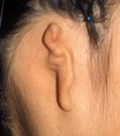
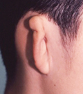
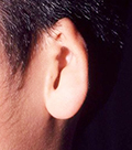
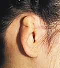
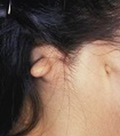
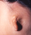
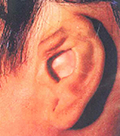
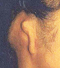
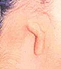
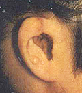
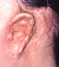
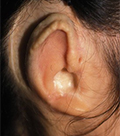
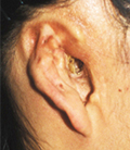
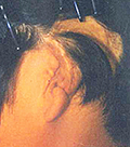
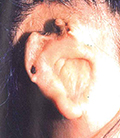
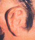
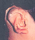
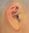
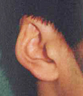
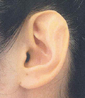
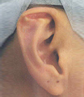
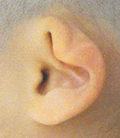
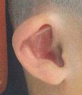
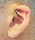
Congenital auricular defects can be classified into: [1] lobule type microtia; [2] small concha type microtia; [3] concha type microtia; [4] anotia and [5-8] those with low hairline. The first stage operation is the fabrication and grafting of the three-dimensional costal cartilage framework [3-D frame]. The second stage operation is the projection of the reconstructed auricle, symmetrically to the contralateral auricle. In both stages, chest wall deformity must be avoided. Recent advancements in auricular reconstruction enabled us to treat complicated cases such as secondary auricular reconstruction, anotia [including traumatic amputation], low hairline cases, hemifacial macrosomia and post-ENT surgery with consistent and more than satisfactory results. The key is to apply the systematic approach of using temporoparietal fascia flap [TPF], deep temporal flap [DTF] and ultra-delicate split- thickness scalp skin [UDSTS]. The approach can be applied to all microtia cases. TPF with UDSTS cover or DTF with UDSTS cover will both function like a skin flap with full vascularity. Thus, there is no postoperative contracture of the skin cover that leads to resorption of the grafted costal cartilage framework. The conventional method of skin grafting alone is contraindicated because it fails to maintain the contour of the auricle, due to vascular compromise, contracture and resorption.


Photos in this blog are shown for your reference for understanding the treatment of microtia. Please understand that surgery results vary depending on each case.
Possible complications following microtia reconstruction surgery
Transient facial palsy, Failure of skin graft due to vascular compromise, Infection resulting in exposure of the 3-D frame, Pneumothorax, Postoperative pneumonia, Suture failure, Alopecia, Decubitus and Others
In the event such complications arise, appropriate action is taken based on the case. Additional or secondary surgery may be required in some situations.


Photos in this blog are shown for your reference for understanding ear reconstruction surgery. Please understand that surgery results vary depending on each case.
Possible complications following ear reconstruction surgery
Infection, Suture failure and Others
In the event such complications arise, appropriate action is taken based on the case. Additional or secondary surgery may be required in some situations.